TL;DR
Your sonographer said "anterior placenta" at your anatomy scan, you opened a search engine, and now you're reading this. Here is what you need to know in three sentences:
- An anterior placenta is normal. It means the placenta attached to the front wall of your uterus, between you and your baby. About half of pregnancies have an anterior placenta and the other half have it on the back, top, or sides — none of these positions are inherently better or worse for the baby.
- It is NOT the same as placenta previa. Placenta previa is a different condition where the placenta covers the cervix; that one has medical implications. Anterior placenta on its own does not.
- The main practical difference is that you may feel your baby's movements a few weeks later than friends do, because the placenta is between the baby's kicks and your belly. That's the main thing. Almost everything else in your pregnancy proceeds normally.
If that's all you came for, you can stop here. The rest of this post is the longer version, plus a few practical things that DO change with an anterior placenta — particularly around 3D ultrasound photo quality, which we'll cover honestly because that's our area of expertise.
What is the placenta, exactly?
The placenta is a temporary organ your body grows specifically for the pregnancy. It attaches to the wall of the uterus, connects to the baby through the umbilical cord, and acts as the baby's life support system: it delivers oxygen and nutrients from your blood, removes waste, and produces hormones that maintain the pregnancy.
It is one of the few organs the human body grows from scratch in adulthood, and it's one of the few that's discarded after use. After the baby is born, the placenta is delivered shortly after (which is why labor has "stages" — Stage 3 is the placenta delivery).
Where the placenta attaches inside the uterus is somewhat random. The uterus has four general regions where it can implant:
| Position | Where the placenta is |
|---|---|
| Anterior | Front wall of uterus (between baby and your belly) |
| Posterior | Back wall of uterus (between baby and your spine) |
| Fundal | Top of uterus |
| Lateral | Right or left side wall |
A placenta can also be a combination — for example "anterior-fundal" means it's mostly on the front wall but extending to the top.
There's no "correct" position. The fertilized egg implants wherever it happens to land in the uterine wall, and the placenta grows from that implantation site.
How common is anterior placenta?
Roughly 50% of pregnancies have an anterior placenta. The exact percentage varies between studies (some put it at 33%, others at 55%, depending on how they categorize combined positions like "anterior-fundal"), but the takeaway is the same: this is one of the two most common placenta positions, alongside posterior.
If your sonographer's tone made it sound like a Big Discovery, that's because they have to mention it. It changes how they conduct the scan (more on that below), but it does not change the medical assessment of your pregnancy on its own.
Anterior placenta vs placenta previa: the critical distinction
Most of the anxiety around the words "anterior placenta" comes from confusing it with placenta previa. They are very different things.
| Anterior placenta | Placenta previa | |
|---|---|---|
| What it is | Placenta on the front wall | Placenta covering the cervix (regardless of front/back) |
| How common | ~50% of pregnancies | ~0.5% of pregnancies (1 in 200) |
| Medical concern | Generally none | Yes — risk of bleeding + may require C-section |
| Detection | Routine — anatomy scan | Routine — anatomy scan |
| Often resolves on its own | N/A (it's normal) | Yes, ~90% resolve as uterus grows |
If your sonographer or OB said "anterior placenta," they did NOT say "placenta previa." These are diagnosed and labeled separately. If there was any concern about previa, they would have used that exact word.
If you're not sure what was said, call your OB's office and ask the nurse to read you the radiologist's report. The terminology is precise and the difference matters.
What anterior placenta DOES change in your pregnancy
Three real, mostly-mild differences:
1. You may feel fetal movement later
The placenta sits between the baby's kicks and your abdominal wall. With an anterior placenta, that "kick" has more cushion to travel through before you feel it. Most moms with an anterior placenta start feeling distinct movements at 20–24 weeks instead of the typical 16–20 weeks for posterior placenta.
This is the single most-asked question about anterior placenta. Your baby is moving normally — you just can't feel it as well yet. By 28–30 weeks, most anterior-placenta moms feel kicks clearly because the baby is bigger and the kicks are stronger.
2. Doppler heartbeat at home may be harder to find
If you bought a home Doppler (which obstetricians generally discourage — see FDA's position), you may have a harder time locating the heartbeat. The Doppler signal has to travel through the placenta, which dampens it. This is not a sign of a problem — it's a sign of physics.
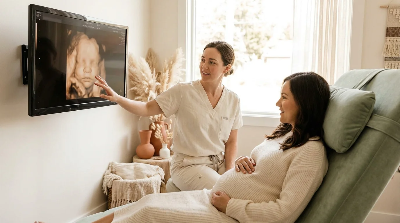
3. 3D ultrasound photos can be harder to capture
This is our area of expertise, so we'll be honest. An anterior placenta sits between the probe and the baby's face during a 3D scan. To get a clean 3D rendering, the sonographer needs a clear pocket of amniotic fluid between probe and face. With an anterior placenta, that pocket is sometimes blocked.
Practical implications for keepsake studios:
- Aim for 26–28 weeks rather than the late end of the sweet spot (30–32 weeks). Earlier in the window, the baby has more room to move, so the chance of finding a good angle around the placenta is better.
- Hydrate aggressively in the 24–48 hours before. More amniotic fluid = more space.
- Choose a studio with a free re-do policy if your first attempt doesn't produce great photos. About 60% of US keepsake studios offer this.
- Allow longer session time. Standard sessions run 15 minutes; some studios offer extended 30-minute sessions (sometimes for an upcharge) which gives the sonographer more time to find a good angle.
We cover all of this in more depth in Best week for a 3D / 4D ultrasound.
The 60% re-do figure comes from intake calls, not website copy — only 1 studio out of the 208 in our audit advertises a free re-do in their public description, which tells you something about what's worth asking on the phone versus what shows up on the homepage. Los Angeles has the deepest market we cover (22 priced 3D studios), so anterior-placenta moms there have the most leverage to compare re-do terms. Smaller markets, ask twice before paying.
What anterior placenta does NOT change
Honest list, since this is what most moms are actually worried about:
- Risk of miscarriage — not increased
- Risk of preterm labor — not increased
- Birth method — generally unchanged (anterior placenta alone does not require C-section)
- Baby's health and development — not affected
- Length of pregnancy — not affected
- Type of prenatal care needed — same as any other pregnancy
- Whether you can feel the baby's hiccups — usually still felt, just slightly muffled
If your OB has specifically flagged a concern (placenta accreta, low-lying placenta, marginal previa), those are separate diagnoses with separate names — they're not the default assumption when "anterior placenta" is mentioned.
When anterior placenta DOES become medically relevant
Three combinations to know about. None of these is "anterior placenta itself caused this" — they're situations where the position becomes relevant to a separate clinical question:
If you've had a previous C-section — there's a small increased risk of placenta accreta (the placenta growing too deeply into the uterine wall) when an anterior placenta sits over a prior C-section scar. Your OB will know about this and may order an additional MRI or specialist ultrasound. (ACOG Committee Opinion 590 covers the full clinical context.)
If amniocentesis is planned — the doctor will plan around the placenta location (anterior or otherwise) to avoid going through it. Anterior placenta doesn't prevent amniocentesis; it just changes the approach.
If a low-lying placenta is also flagged — "low-lying anterior" is a phrase that means the placenta is anterior AND extends close to the cervix. This usually resolves as the uterus grows in the third trimester, but your OB will recheck around 28–32 weeks.
In all three cases, your OB will explicitly tell you. If they didn't, none of these situations apply to you.
Will the placenta move?
Yes, and no.
The placenta itself doesn't actively migrate, but the uterus grows during pregnancy. As the uterus expands, the placenta gets carried along — and what looks like a low-lying placenta at 20 weeks can sit comfortably away from the cervix by 32 weeks.
This is why your OB doesn't make decisions about a low-lying placenta until later in pregnancy. About 90% of placenta previa diagnoses at 18–22 weeks resolve to "no longer covering cervix" by 32–34 weeks, just from the uterus growing upward and outward.
The position relative to the front/back/top/sides of the uterus doesn't usually change — anterior tends to stay anterior. But the specific question of whether it's near the cervix is dynamic and gets re-checked.
Practical tips for the rest of your pregnancy
If you've been told you have an anterior placenta and you don't have any other complicating factor:
Be patient about feeling movement. Compared to friends with posterior placenta, you may not feel distinct kicks until 22–24 weeks. This is normal. Once you do start feeling them, you'll feel them clearly.
Use kick counts in the third trimester. Your OB will instruct you on tracking baby movements daily after 28 weeks. The methodology is the same regardless of placenta position. Anterior moms tend to feel a slightly different "shape" of movement (more rolls and stretches than sharp kicks), but the count requirement is the same.
Plan a 3D session for week 26–28 (if you want one). Earlier is better with anterior placenta. See best week for a 3D ultrasound and 3D ultrasound cost for the timing and pricing context.
Don't buy a home Doppler. The FDA explicitly recommends against home fetal heartbeat monitors for any pregnancy, and they're particularly unreliable with an anterior placenta. If you have a concern about movement, call your OB's nurse line. That's what they're there for.
At the 28–32 week recheck, your OB or sonographer will reconfirm placenta position. If the position is still anterior (and not low-lying), no further action.
Frequently asked questions
Is anterior placenta high risk?
No. Anterior placenta on its own is not high-risk. It is a normal anatomical variation present in roughly half of pregnancies. High-risk conditions involving the placenta — placenta previa, placenta accreta, vasa previa — are diagnosed by name; they are not assumed from "anterior placenta."
Why didn't my doctor mention anterior placenta?
Most OBs don't lead with placenta position unless there's a clinical reason. The radiologist's report from your anatomy scan will note it, but unless it requires a change to your care plan, your OB may not bring it up. If you want to know your placenta position, you can ask at your next appointment — it's documented in your record.
Does anterior placenta affect baby's growth?
No. The placenta's nutrient and oxygen exchange function works the same whether it's attached to the front, back, top, or sides of the uterus. Placenta dysfunction is a separate clinical issue (intrauterine growth restriction, etc.) and is monitored by ultrasound regardless of position.
Can I have a vaginal birth with anterior placenta?
Yes, in nearly all cases. Anterior placenta is not a reason for C-section. The only placenta-position reason for planned C-section is placenta previa (placenta covering the cervix), which is a separate diagnosis.
Does anterior placenta increase risk of bleeding?
Not by itself. Bleeding risk increases with placenta previa, placenta accreta, and a few other specific conditions, none of which are implied by "anterior" alone.
Will anterior placenta show up on every ultrasound?
The placenta is described in the radiologist's report at every detailed ultrasound (anatomy scan and any growth scans). At quick OB office scans for heartbeat or position checks, it may not be documented because it isn't relevant to the question being asked.
Can I feel baby's kicks with anterior placenta?
Yes — eventually. Most anterior-placenta moms start feeling distinct movement around week 22 to 24 (versus 16 to 20 weeks for posterior). By week 28, kick perception is comparable to other pregnancies because the baby is bigger and the kicks are stronger.
Does anterior placenta affect 3D ultrasound photo quality?
Sometimes, yes. The placenta sits between the probe and the baby's face, which can block the clear amniotic fluid pocket needed for 3D rendering. Booking earlier in the 26–32 week sweet spot, hydrating well, and choosing a studio with a free re-do policy help. See our best week for 3D ultrasound for the full guide.
Do twins with anterior placentas have the same situation?
Twin pregnancies have their own placenta logistics depending on whether they share a placenta (monochorionic) or have separate ones (dichorionic). Anterior position in twin pregnancies is even more common because there's more uterine surface used. Your OB or maternal-fetal medicine specialist will speak to your specific case.
If your concern was simply "what does this mean," the answer is: not much, and you can keep doing what you've been doing. If you wanted to know how it might affect a planned 3D ultrasound, our best week for 3D ultrasound and verified studio directory cover the practical decisions. And if you have specific concerns about your pregnancy, your OB's nurse line is the right place to ask — not a search engine.